Essential Aspects to Check in Optical Biometry to Avoid Refractive Surprises Post IOL Implantation
- Subhabrata Bhattacharya

- 20 hours ago
- 7 min read
Updated: 3 hours ago
Optical biometry has transformed the way ophthalmologists calculate intraocular lens (IOL) power before cataract surgery. Despite advances, refractive surprises—unexpected vision outcomes after IOL implantation—still occur. These refractive surprises can lead to patient dissatisfaction and sometimes require additional procedures. To minimize this risk, it is crucial to carefully analyze the optical biometry chart and ensure all key parameters are accurate and reliable before you jump to your favorite IOL calculation formula.
This post highlights ten essential aspects to check in the optical biometry chart by way of which you can largely avoid refractive surprise. Understanding these factors helps surgeons make informed decisions, improving refractive outcomes and patient satisfaction.

1. Validating the measurements - A measurement is only as good as our ability to validate the data generated
Before you accept the patient's optical biometry measurements, validate that the measurements can be relied upon.
Always do a two eye biometry. This would help you to corelate the two eye measurements and rule out abnormality, that may need remeasurement.

For Tomey OA-2000 or IOL Master keep an eye on the SNR values. For axial length measurements on the ZEISS IOL Master 500, the Signal-to-Noise Ratio (SNR) indicates the quality and reliability of the interferometric peak used for axial length calculation.
Typical interpretation of IOL Master 500 SNR values:
SNR | Interpretation | Clinical meaning |
<2.0 | Poor | Unreliable; repeat scan or do an immersion biometry |
2-5 | Borderline | Use cautiously; repeat if possible, corelate with immersion biometry |
>5 | Good quality | Reliable |
>10 | Very good quality | High confidence |
OA- 2000 generate tomographic images of retina that can be useful in data validation.
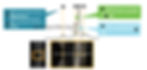
A good reading with Tomey OS-2000 should have the following features:
Distinct peaks depicting B ellipsoid zone, C Retina and D sclera. Additionally, ILM membrane (A) may be visible
Additionally, the SNR value should be higher than 3.

2. Corneal Curvature (Keratometry) Values

Keratometry measures the corneal curvature, which affects the eye’s refractive power. Ensure the following:
The difference of astigmatism is less than 1 diopter between the two eyes.
The corneal spherical equivalent (SE) difference between both eyes is less than 1 diopter.
Too steep a cornea (>47D) or too flat a cornea (<41D) may need re measurement or validated with topography to rule out irregularity. In the image 4b, a way out of the whack value of 49.12 diopters in the right eye should raise suspicion and be followed up with corneal topography.

Too low an astigmatism (<1 diopter) with high standard deviation of K readings may indicate dry eyes.
In the Tomey OA-2000, check for KRI (corneal regularity/irregularity index) and KAI (corneal asymmetry index values).
KAI | 0 - 23.4 |
KRI | 0- 4.4 |
KAI and KRI values should be between the above values to indicate a healthy cornea quickguide.org | |
3. Axial Length (AXL values)
Every .1 mm of error in axial length measurement in an average eye, can lead to .3 diopter of difference in refractive error.
Therefore, the difference in axial length of .3 mm or more in an average to long eye, should be rechecked.
For short eyes, or axial hyperopia, consider remeasurement when the difference in axial length between the two eyes are equal to or more than .2 mm.

4. Standard Deviation of Axial Length and Corneal Measurement values
Following are the recommended values for standard deviation.

For optical biometry like IOL Master, the SD of AXL values should be less than .05 mm.
For optical biometry, the SD of K readings should be <.20 diopter
The SD of axis for K readings should be <3.5 deg.

Image 7: Standard deviation value displayed for axial length in the Tomey quickguide.org
5. No Drops Before Biometry
It is important to do IOL power calculation and biometry before applying any drop on the cornea. This is because, applying drops on the cornea may lead to steeper values of cornea, that may lead to a lower IOL power and thereby lead to a hyperopic surprise.

6. Do a Bscan for high axial length patients.
Patients with high axial myopia, may be associated with posterior staphyloma. The incidence of posterior staphyloma increases in patients with high axial myopia, particularly beyond 27.0 mm of axial length.


In posterior staphyloma, the anatomical axial length and the refractive axial length do not match. The anatomical axial length is higher than the refractive axial length, which depicts the distance from the cornea to the fovea. As a result, often a higher axial length is landed up with in ultrasound biometry. In optical biometry, if the patient has not fixated on the light, chances are that the measurement is not on the fovea. Therefore, a Bscan should be done to validate the posterior vitreous length. Note, the fovea is situated around 5-6 mm away from the optic nerve, that would help locate the fovea in the BScan (Image 10).
7. Presence of Ocular Surface Abnormalities

Dry eye, corneal scars, or irregularities can affect measurement accuracy. No optical biometry or auto keratometry device can tell you that the patient has irregular astigmatism. These devices hypothesize that the flat meridian is always 90 degrees away from the steep meridian. Therefore, other than the points stated in Section 2 with regard to corneal values, keep in mind the following:
Low astigmatism ( less than 1 diopter) with high standard deviation of magnitude (>.2 diopter) and axis (>3.5 deg) may indicate dry eye.
Treat dry eye or other surface problems before biometry to improve data quality.
If a topography is available, corelate measurement with topography.

Dry eyes can result from aqueous deficiency involving lacrimal glands, or evaporative (meibomian gland dysfunction) or a combination of both. While Schirmer's test is common in a clinic, it does not throw light if there is an underlying MGD involved.
Thus a comprehensive examination of ocular abnormalities need to be checked before a premium IOL is planned.
8. For premium IOLs like multi focal, corelate the pupillary diameter of two eyes
The difference of pupil diameter of two eyes should be less than 1 mm. A difference of more than 1 mm diameter of pupil of both eyes could indicate pathologic anisocoria. Thus light and dark room evaluation should follow before a premium IOL is implanted.
If the anisocoria is more in dark condition, the constricted pupil may be pathologic
If the anisocoria is more in bright light condition, the dilated pupil may be pathologic.
If the anisocoria is same in both light and dark conditions, it may be indicative of physiologic anisocoria.
For more information, do follow my article on pupillary evaluation https://www.quickguide.org/post/pupil-test
9. For premium IOLs, check for HOA, and large decentration of eye
Three most important higher order aberrations that lead to a drop in image quality and photic phenomenon with diffractive multifocal IOLs are the spherical aberration, coma and trefoil.
Total HOA RMS value of more than .5 micron may be a red or at least a yellow flag.
Individual values of spherical aberration, coma and trefoil may be less than .3 micron
Angle Kappa and Angle Alpha in optical biometry machines are displayed often by x/y coordinates. Where chord mu or Chang-Waring (CWC) chord values are not provided, you may add the x/y co-ordinate values and exclude patients with more than .5 mm of decentration.
For CWC and chord mu values in IOL Master, use a value of less than .4 mm
For angle kappa and alpha values in iTrace, follow the yellow, green indication.

Image 12: Follow the X/Y coordinate values to rule out major decentration in the eye. PCX and PCY indicates decentration of pupil barycenter from corneal vertex/visual axis quickguide.org
In Image 12, Iris barycenter and pupil barycenter in terms of X/Y coordinates are throwing information with respect to decentration (chord distance) from the corneal vertex (close to visual axis). To understand more you can follow the below link that describes the concept:
Understanding Angle Alpha & Angle Kappa- the finer aspects
An excerpt from the article is provided here below:

10. A constant optimization
If a constant personalization is impossible in a busy practice, follow iolcon.org as a starting point for IOL constants for any new IOL wherever applicable. Personalization is recommended after at least 30 cases with same IOL, incision and capsulorhexis size. Personalization of A constant with a particular IOL is recommended particularly for short eyes. For such patients, keep a record of outcome, for axial length below 23 mm and corneal curvature above 45 diopters.
To help you with quick optimization, here is a simple optimization tool that you can follow:

By carefully reviewing these ten aspects in the optical biometry chart, surgeons can significantly reduce the risk of refractive surprises after IOL implantation. Accurate measurements, consistent data, and appropriate formula selection form the foundation of successful cataract surgery outcomes.
Taking the time to verify each parameter and understanding its impact on IOL power calculation leads to better vision results and higher patient satisfaction. If any measurement appears questionable, repeating the scan or using alternative methods can prevent costly postoperative corrections.


